Hyper-wide-angle fundus camera allowing wide field fundus photography
Joint development with Nara Institute of Science and Technology
-

-
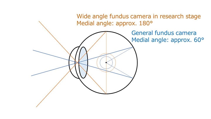
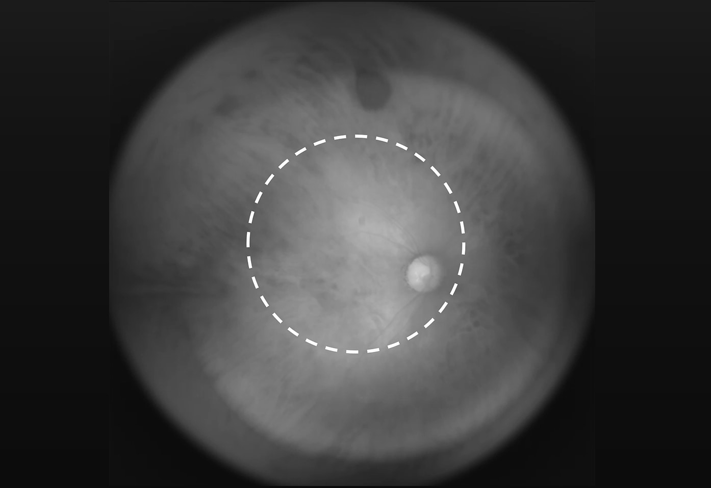
Fundus camera are used for early detection of frequent causes in Japan of blindness such as glaucoma, retinitis pigmentosa and diabetic retinopathy. Although wider view angle is necessary for eye fundus imaging and examination, existing cameras currently used for medical examination have viewing angle of 60°which detects only part of eye fundus. There are difficulties in designing a lens for clear photography of a concave sphere such as the fundus, and projecting light through the lens to the fundus. Tamron has specifically designed and completed a prototype of hyper-wide-angle lens optimized for fundus photography. The lens was installed into near-infrared fundus camera developed by Nara Institute of Science and Technology and approximately 180° of hyper-wide viewing angle photographing was achieved. Without mydriatic (eye drop) used to dilate the pupils, a wide field of eye fundus image can be acquired.
- Tamron’s prototype lens with compact and hyper-wide-angle optimized for fundus photography
- Various technologies illuminating stably near infrared through pupil to wide fundus, which are newly developed by Nara Institute of Science and Technology
- Hyper-wide-angle (180°) fundus photography was achieved by combining the two technologies
- Wide field of eye fundus image can be acquired without mydriatic (eye drop)

Pattern diagram of available imaging field

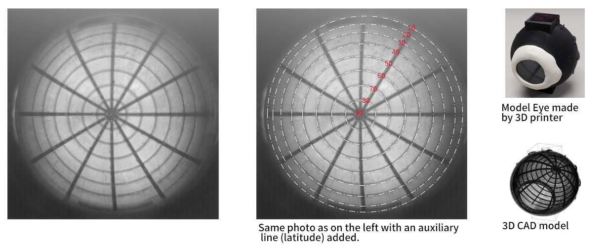
View field checking image by 3D printer model eye
Opto-Science R&D
development
20230215002947

